Nov 23, 2008

Poxviridae

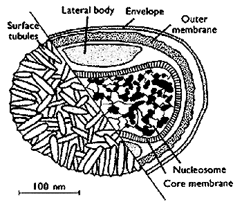
In today’s lecture, I learnt about this very deadly virus in the past that killed lots of people, which is extinct already. It is called, “small pox”. It comes from a family called, poxviridate, which is the largest family of viruses, and can be seen under a microscope. But be careful, it is deadly as it can remain stable for hours in air allowing it to spread easily!

clip_image001

http://www.stanford.edu/group/virus/pox/2000/IMAGES/poxtoon.gif

Its genome is double stranded DNA, covalently cross-linked at ends.

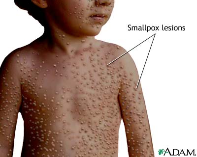
Small pox infects human only. It causes respiratory secretions, and is associated with skin lesions.

clip_image002

http://www.nlm.nih.gov/medlineplus/ency/images/ency/fullsize/19059.jpg

It symptoms is initially influenza-like. It will result in neurological damage, blindness, and even death.

Variola major and Variola minor are variants of small pox.

Some statistics of these variants were gathered in the lecture:

Variola Major – 25 to 30% fatalities

Variola Minor – less than 1% death

Now, let me tell you about vaccination that helped in smallpox eradication.


http://www.ebinrushed.com/history/images/history_7.jpg

It was found by Edward Jenner who observed that milkmaids got cowpox virus, also known as vaccinia, from cow, and didn't seem to get very sick when infected with small pox and always recovered. So, he decided to test out his theory, he vaccinated an 8 year old boy, by the name of James Phipps. There after, he was challenged with small pox, and the similar effects were observed - the symptoms of small pox were milder and recovery was quick.

From this virus, the term VACCINATION was coined. The derivative of vaccinia, the cowpox virus.

With this new weapon called vaccination, and people obtaining lifelong immunity after recovering from small pox, and human being its only reservoir to infect, the smallpox virus is finally eradicated.

It was announced to be eradicated by WHO (World Health Organisatio) in 1980! What an exciteful lecture on VIROLOGY!
Nov 14, 2008

Retroviridae

http://www.stanford.edu/group/virus/retro/2005gongishmail/hiv1.jpg

Week 5 Lecture on Retroviruses was not bad. What is retrovirus? A retrovirus is a virus with an RNA genome that replicates by using a viral reverse transcriptase enzyme to transcribe its RNA into DNA in the host cell. The lecture was mostly about Human T-lymphotropic virus (HTLV-1) and Human Immunodeficiency virus (HIV). We also learned about the history and characteristics of retroviruses. Did you know that the first human retrovirus was discovered in 1981? It was the Human T-lymphotropic virus. Retroviruses have received much attention even before 1980. It had a long history that dates back
to 1908. That is almost 100 years ago.
  • 1908: Ellerman and Bang, searching for an infectious cause for leukaemia, studied leukaemia in chickens and succeed in transferring the disease from one to another by cell-free tissue filtrates
  • 1911: Peyton Rous discovered chicken sarcoma caused by a virus
  • 1960’s: Howard Temin knew that retrovirus genomes were composed of RNA and observed that replication was inhibited by actinomycin D which inhibits DNA synthesis therefore he proposed the concept of reverse transcription.
  • 1970:Michael Bishop and Harold Varmus discovered oncogenes and role of oncogenic viruses and cancer
  • 1980: Human T-cell leukaemia virus discovered. The first pathogenic human retrovirus.
  • 1983: Human immunodeficiency virus discovered.

Retroviridae are broken down further into the genus as show in the table below:

Genus

Examples

Alpharetrovirus

Avian leukosis virus

Betaretrovirus

Mouse mammary tumor virus

Gammaretrovirus

Murine leukernia virus

Deltaretrovirus

Human T-lymphotropic virus

Epsilonretrovirus

Walley dermal sarcoma virus

Lentivirus

Human immunodeficiency virus

Spumavirus

Human foamy virus

The life cycle of retroviruses:

image

http://www.gene.com/AE/AB/GG/examples_of_viruses.html

Main steps are Attachment, Entry, Uncoating, Transcription, Translation, Assembly and Release.

HTLV-1

What is HTLV-1? It’s the virus that causes Adult T-cell leukaemia (ATLL) which is a rare cancer to the T-cells. Acute aggressive leukaemia can kill in 12 months. Unfortunately, there is no known cure yet.

HIV

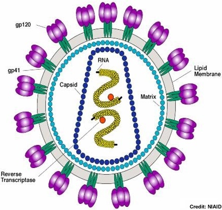
HIV is the virus that cause acquired immunodeficiency syndrome (AIDS), a condition in humans in which the immune system begins to fail, leading to life-threatening opportunistic infections.The picture below show the life cycle and structure of HIV:

hiv

http://commons.wikimedia.org/wiki/File:Hiv_gross.png

HIV can be transmitted through sexual contact, blood or from mother to child. Symptom of a HIV infection are:

During Primary infection:

  • acute stage
  • flu-like symptoms
  • fever
  • skin rash
  • swollen lymph nodes

During Asymptomatic stage:

  • fatigue
  • depression
  • weight loss
  • memory disorders

Another deadly virus, yet no cure is available. However a lot of research is in progress. Possible methods is vaccines or to inhibit any of the stages of the HIV life cycle.

Nov 13, 2008

Flaviridae

During week five, the topic on Flaviviridae was taught during lecture time. Let me give you more details about this particular virus.

Baltimore Classification

Viruses; ssRNA positive-strand viruses, no DNA stage; Flaviviridae

Description and Significance

  • Flaviviridae contains a myriad of viruses that cause disease in humans. Flaviviridae has a total of 69 pathogens in its rank. The family gets its name from Yellow Fever virus, a type virus of Flaviviridae; flavus means yellow in Latin.
  • The Flavivirus genus contains several nasty critters that include yellow fever virus, dengue fever virus, and Japanese encephalitis (JE) virus.

Morphology

  • Virions uniform in shape; spheroidal; enveloped; 40-60 nm in diameter. Surface projections of envelope small (surface appears rough), or distinct (and are obvious fringes in negative stains). Nucleocapsids isometric; 25-30 nm in diameter. Symmetry polyhedral.

Major diseases caused by the Flaviviridae family include:

  • Dengue fever
  • Japanese encephalitis
  • Kyasanur Forest disease

Dengue Fever

http://www.stanford.edu/group/virus/flavi/2000/mosquito_mov.gif
  • Dengue Fever and Dengue Haemorrhagic Fever (a more severe form) are the most common mosquito-borne viral diseases in the world.
  • The diagnosis of dengue is usually made clinically. The classic picture is high fever with no localising source of infection, a petechial rash with thrombocytopenia and relative leukopenia - low platelet and white blood cell count.
  • Signs and symptoms include a sudden onset of severe headache, muscle and joint pains, fever, and rash.
  • The classic dengue fever lasts about six to seven days, with a smaller peak of fever at the trailing end of the disease (the so-called biphasic pattern). Dengue Vaccine Initiative which was set up in 2003 with the aim of accelerating the development and introduction of dengue vaccine(s) that are affordable and accessible to poor children in endemic countries.

Japanese encephalitis

  • Japanese encephalitis is a disease caused by the mosquito-borne Japanese encephalitis virus. The Japanese encephalitis virus is a virus from the family Flaviviridae.
  • Domestic pigs and wild birds are reservoirs of the virus; transmission to humans may cause severe symptoms. One of the most important vectors of this disease is the mosquito Culex tritaeniorhynchus. This disease is most prevalent in Southeast Asia and the Far East.
  • Japanese encephalitis has an incubation period of 5 to 15 days and the vast majority of infections are asymptomatic: only 1 in 250 infections develop into encephalitis. There is no specific treatment for Japanese encephalitis and treatment is supportive.

The use of arctigenin has been shown to be effective in a mouse model of Japanese encephalitis, but there is as yet no clinical evidence to support its use.

Nov 6, 2008

RNA viruses -Orthomyxoviridae & Picornaviridae

All living things have DNA as genetic material. Only viruses have RNA as its genetic material! RNA virus’s genome is highly infectious(+RNA viruses) as it can be read as mRNA directly.

All RNA virus’s have unusual structural characteristics (leader sequence) and can be differentiated between viral mRNA and host mRNA.

Two types of RNA viruses

Orthomyxoviridae and Picornaviridae

Picornaviridae also known as common cold

http://education.expasy.org/images/Picornaviridae_virion.jpg

Morphology(shape)

Virions consist of a capsid. Virus capsid is not enveloped, round with icosahedral symmetry. The capsid is isometricand and capsids appear round. The capsid consists of 12 capsomers.

Incomplete particles are common.

Genome

The genomic RNA of picornaviridae is infectious. It is long untranslated region end of 5’ end “clover leaf” structure (IRES). It is one linear (+) RNA.

Single polyprotein of 2100 –2400 aa

Both of the ends are modified.

Pathogenesis

Upper Respiratory Tract infection

Short Incubation ( 2 –3 days)

Endogenous IFN helps!

Locally synthesized IgA but titer decline with time

So what are the symptoms?

  • Water nasal discharge
  • Sneezing
  • Little/No fever
  • Congestion

When do we get Picornaviridae?

All throughout the year, virus abundant in nasal discharge.

How can we control it?

  • Wash hands
  • Keep hands away from eyes and nose
  • Sneeze into tissue & throw away
  • Maintain personal hygiene
  • Stay away from people who have colds.

Is vaccination possible for Picornaviridae?

Yes. It is possible. However vaccination is not so useful because we get common cold throughout the whole year and so it is not much use. Another reason is that vaccination is very expensive. So vaccination is not that useful.

Orthomyxoviridae also known as Influenza

http://www.pasteur.ac.ir/researchDepartment/flu/images/flu_structure.gif

Morphology(shape)

Typically Spherical (100nm diameter)

  • Enveloped ,Pleomorphic (many forms)
  • Spikes on envelope
  • Groups of HA (hemagglutinin) or NA (neuraminidase)
  • Ratio of HA to NA
    • 5:1

Genome

ss (-) RNA in 8 segments

  • 3 polymerase polypeptides with each segment
  • 5’ and 3’ end of all segments highly conserved

Pathogenesis

  • Respiratory Tract in human
  • Affinity of the HA for receptors in the epithelium of the tract
  • Innate resistance
  • Mucus blanket/cilia
  • mmunocompromised

Lab Diagnosis

  • Throat swab/gargle/nasal wash/
  • Virus culture (MDCK cell line or chick embryo)

Epidemiology

  • All throughout the year!
  • Incubation (1 –4 days)
  • A few serotypes circulating simultaneously

Differences between Flu and Cold

Symptoms

Cold

Flu

Fever

Rare

Characteristic, high (100-102 degrees F); lasts 3-4 days

Headache

Rare

Prominent

General Aches, Pains

Slight

Usual; often severe

Fatigue, Weakness

Quite mild

Can last up to 2-3 weeks

Extreme Exhaustion

Never

Early and prominent

Stuffy Nose

Common

Sometimes

Sneezing

Usual

Sometimes

Sore Throat

Common

Sometimes

Chest Discomfort,
Cough

Mild to moderate;
hacking cough

Common; can become severe

Complications

Sinus congestion or earache

Bronchitis, pneumonia; can be life-threatening

Prevention

Good hygiene

Annual flu shot or FluMist

Treatment

Only temporary relief of symptoms

Antiviral drugs (oseltamivir or zanamavir) within 24-48 hours of onset